

Nanoengineers at the University of California, San Diego have developed a novel technology that can fabricate, in mere seconds, microscale three dimensional (3D) structures out of soft, biocompatible hydrogels. Near term, the technology could lead to better systems for growing and studying cells, including stem cells, in the laboratory. Long-term, the goal is to be able to print biological tissues for regenerative medicine. For example, in the future, doctors may repair the damage caused by heart attack by replacing it with tissue that rolled off of a printer.
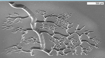
NanoEngineering Professor Shaochen Chen has demonstrated the capability of printing three-dimensional blood vessels in mere seconds out of soft, biocompatible hydrogels. Being able to print blood vessels is essential to achieving the promise of regenerative medicine because it is how the body distributes oxygen and nutrients. Image Credit: Biomedical Nanotechnology Laboratory, Chen Research Group, UC San Diego Jacobs School of Engineering.
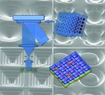
The biofabrication technology, called dynamic optical projection stereolithography (DOPsL), was developed in the laboratory of NanoEngineering Professor Shaochen Chen. Current fabrication techniques, such as photolithography and micro-contact printing, are limited to generating simple geometries or 2D patterns. Stereolithography is best known for its ability to print large objects such as tools and car parts. The difference, says Chen, is in the micro- and nanoscale resolution required to print tissues that mimic nature’s fine-grained details, including blood vessels, which are essential for distributing nutrients and oxygen throughout the body. Without the ability to print vasculature, an engineered liver or kidney, for example, is useless in regenerative medicine. With DOPsL, Chen’s team was able to achieve more complex geometries common in nature such as flowers, spirals and hemispheres. Other current 3D fabrication techniques, such as two-photon photopolymerization, can take hours to fabricate a 3D part.
The biofabrication technique uses a computer projection system and precisely controlled micromirrors to shine light on a selected area of a solution containing photo-sensitive biopolymers and cells. This photo-induced solidification process forms one layer of solid structure at a time, but in a continuous fashion. The technology is part of a new biofabrication technology that Chen is developing under a four-year, $1.5 million grant from the National Institutes of Health (R01EB012597). The Obama administration in March launched a $1 billion investment in advanced manufacturing technologies, including creating the National Additive Manufacturing Innovation Institute with $30 million in federal funding to focus on 3D printing. The term “additive manufacturing” refers to the way 3D structures are built layering very thin materials.
The Chen Research Group is focused on fabrication of nanostructured biomaterials and nanophotonics for biomedical engineering applications and recently moved into the new Structural and Materials Engineering Building, which is bringing nano and structural engineers, medical device labs and visual artists into a collaborative environment under one roof.
ABSTRACT – The topographic features of the extracelluar matrix (ECM) lay the foundation for cellular behavior. A novel biofabrication method using a digital-mirror device (DMD), called dynamic optical projection stereolithography (DOPsL) is demonstrated. This robust and versatile platform can generate complex biomimetic scaffolds within seconds. Such 3D scaffolds have promising potentials for studying cell interactions with microenvironments in vitro and in vivo.
If you liked this article, please give it a quick review on ycombinator or StumbleUpon. Thanks

Brian Wang is a Futurist Thought Leader and a popular Science blogger with 1 million readers per month. His blog Nextbigfuture.com is ranked #1 Science News Blog. It covers many disruptive technology and trends including Space, Robotics, Artificial Intelligence, Medicine, Anti-aging Biotechnology, and Nanotechnology.
Known for identifying cutting edge technologies, he is currently a Co-Founder of a startup and fundraiser for high potential early-stage companies. He is the Head of Research for Allocations for deep technology investments and an Angel Investor at Space Angels.
A frequent speaker at corporations, he has been a TEDx speaker, a Singularity University speaker and guest at numerous interviews for radio and podcasts. He is open to public speaking and advising engagements.