
Bioengineers can already make 2D structures out of many kinds of tissue, but one of the major roadblocks to making the jump to 3D is keeping the cells within large structures from suffocating; organs have complicated 3D blood vessel networks that are still impossible to recreate in the laboratory.
Now, University of Pennsylvania researchers have developed an innovative solution to this perfusion problem: they’ve shown that 3D printed templates of filament networks can be used to rapidly create vasculature and improve the function of engineered living tissues.

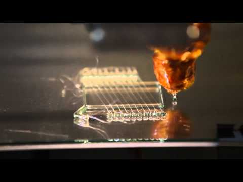
A microscope image of a 3D printed sugar template used for creating vasculature in living tissues. (Photo: Jordan S. Miller)
Without a vascular system — a highway for delivering nutrients and removing waste products — living cells on the inside of a 3D tissue structure quickly die. Thin tissues grown from a few layers of cells don’t have this problem, as all of the cells have direct access to nutrients and oxygen. Bioengineers have therefore explored 3D printing as a way to prototype tissues containing large volumes of living cells.
The most commonly explored techniques are layer-by-layer fabrication, or bioprinting, where single layers or droplets of cells and gel are created and then assembled together one drop at a time, somewhat like building a stack of LEGOs.
Such “additive manufacturing” methods can make complex shapes out of a variety of materials, but vasculature remains a major challenge when printing with cells. Hollow channels made in this way have structural seams running between the layers, and the pressure of fluid pumping through them can push the seams apart. More important, many potentially useful cell types, like liver cells, cannot readily survive the rigors of direct 3D bioprinting.
To get around this problem, Penn researchers turned the printing process inside out.
Rather than trying to print a large volume of tissue and leave hollow channels for vasculature in a layer-by-layer approach, Chen and colleagues focused on the vasculature first and designed free-standing 3D filament networks in the shape of a vascular system that sat inside a mold. As in lost-wax casting, a technique that has been used to make sculptures for thousands of years, the team’s approach allowed for the mold and vascular template to be removed once the cells were added and formed a solid tissue enveloping the filaments.
“Sometimes the simplest solutions come from going back to basics,” Miller said. “I got the first hint at this solution when I visited a Body Worlds exhibit, where you can see plastic casts of free-standing, whole organ vasculature.”
This rapid casting technique hinged on the researchers developing a material that is rigid enough to exist as a 3D network of cylindrical filaments but which can also easily dissolve in water without toxic effects on cells. They also needed to make the material compatible with a 3D printer so they could make reproducible vascular networks orders of magnitude faster, and at larger scale and higher complexity, than possible in a layer-by-layer bioprinting approach.
Monolithic tissue construct containing patterned vascular architectures and living cells. Schematic overview. An open, interconnected, self-supporting carbohydrate-glass lattice is printed to serve as the sacrificial element for the casting of 3D vascular architectures.
After much testing, the team found the perfect mix of material properties in a humble material: sugar. Sugars are mechanically strong and make up the majority of organic biomass on the planet in the form of cellulose, but their building blocks are also typically added and dissolved into nutrient media that help cells grow.
“We tested many different sugar formulations until we were able to optimize all of these characteristics together,” Miller said. “Since there’s no single type of gel that’s going to be optimal for every kind of engineered tissue, we also wanted to develop a sugar formula that would be broadly compatible with any cell type or water-based gel.”
The formula they settled on — a combination of sucrose and glucose along with dextran for structural reinforcement — is printed with a RepRap, an open-source 3D printer with a custom-designed extruder and controlling software. An important step in stabilizing the sugar after printing, templates are coated in a thin layer of a degradable polymer derived from corn. This coating allows the sugar template to be dissolved and to flow out of the gel through the channels they create without inhibiting the solidification of the gel or damaging the growing cells nearby. Once the sugar is removed, the researchers start flowing fluid through the vascular architecture and cells begin to receive nutrients and oxygen similar to the exchange that naturally happens in the body.
The whole process is quick and inexpensive, allowing the researchers to switch with ease between computer simulations and physical models of multiple vascular configurations.
“This new platform technology, from the cell’s perspective, makes tissue formation a gentle and quick journey,” Chen said, “because cells are only exposed to a few minutes of manual pipetting and a single step of being poured into the molds before getting nourished by our vascular network.”
The researchers showed that human blood vessel cells injected throughout the vascular networks spontaneously generated new capillary sprouts to increase the network’s reach, much in the way blood vessels in the body naturally grow. The team then created gels containing primary liver cells to test whether their technique could improve their function.
When the researchers pumped nutrient-rich media through the gel’s template-fashioned vascular system, the entrapped liver cells boosted their production of albumin and urea, natural components of blood and urine, respectively, which are important measures of liver-cell function and health. There was also clear evidence of increased cell survival around the perfused vascular channels.
And theoretical modeling of nutrient transport in these perfused gels showed a striking resemblance to observed cell-survival patterns, opening up the possibility of using live-cell data to refine computer models to better design vascular architectures.
Though these engineered tissues were not equivalent to a fully functioning liver, the researchers used cell densities that approached clinical relevance, suggesting that their printed vascular system could eventually be used to further research in lab-grown organs and organoids.
“The therapeutic window for human-liver therapy is estimated at one to 10 billion functional liver cells,” Bhatia said. “With this work, we’ve brought engineered liver tissues orders of magnitude closer to that goal, but at tens of millions of liver cells per gel we’ve still got a ways to go.
“More work will be needed to learn how to directly connect these types of vascular networks to natural blood vessels while at the same time investigating fundamental interactions between the liver cells and the patterned vasculature. It’s an exciting future ahead.”
With promising indications that their vascular networks will be compatible with all types of cells and gels, the team believes their 3D printing method will be a scalable solution for a wide variety of cell- and tissue-based applications because all organ vasculature follows similar architectural patterns.
“Cell biologists like the idea of 3D printing to make vascularized tissues in principle, but they would need to have an expert in house and highly specialized equipment to even attempt it,” Miller said. “That’s no longer the case; we’ve made these sugar-based vascular templates stable enough to ship to labs around the world.”
Beyond integrating well with the world of tissue engineering, the researchers’ work epitomizes the philosophy that drives much of the open source 3D printing community.
“We launched this project from innovations rooted in RepRap and MakerBot technology and their supporting worldwide communities,” Miller said. “A RepRap 3D printer is a tiny fraction of the cost of commercial 3D printers, and, more important, its open-source nature means you can freely modify it. Many of our additions to the project are already in the wild.”
Several of the custom parts of the RepRap printer the researchers used to make the vascular templates were printed in plastic on another RepRap. Miller will teach a class on building and using these types of printers at a workshop this summer and will continue tinkering with his own designs.
“We want to redesign the printer from scratch and focus it entirely on cell biology, tissue engineering and regenerative medicine applications,” Miller said.

6 pages of supplemental material
If you liked this article, please give it a quick review on ycombinator or StumbleUpon. Thanks

Brian Wang is a Futurist Thought Leader and a popular Science blogger with 1 million readers per month. His blog Nextbigfuture.com is ranked #1 Science News Blog. It covers many disruptive technology and trends including Space, Robotics, Artificial Intelligence, Medicine, Anti-aging Biotechnology, and Nanotechnology.
Known for identifying cutting edge technologies, he is currently a Co-Founder of a startup and fundraiser for high potential early-stage companies. He is the Head of Research for Allocations for deep technology investments and an Angel Investor at Space Angels.
A frequent speaker at corporations, he has been a TEDx speaker, a Singularity University speaker and guest at numerous interviews for radio and podcasts. He is open to public speaking and advising engagements.