

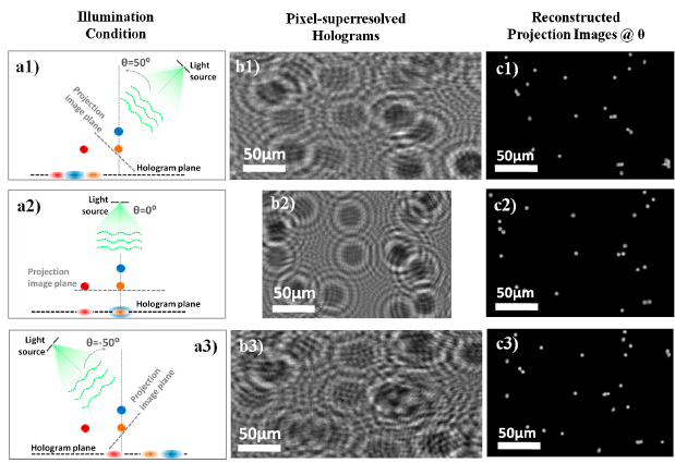
(A1–A3) Schematic illustration of the holographic
recording condition for three angles, 50°, 0°, and −50°, respectively. (B1–B3) Cropped images from corresponding superresolved holograms of 5 μm beads (at z ¼ ∼0.8 mm) measured at illumination angles shown in A1–A3. The holograms of individual beads have an elliptical shape, as expected, since detection plane is not normal to beam propagation. (C1–C3) Digitally reconstructed lens-free projection images using the corresponding holograms in B1–B3. After perspective correction (see SI Text), the ellipticity is removed as revealed by the circular shape of the reconstructed beads. The reconstructed projection images are registered with respect to the bead at the center of the images, which is assumed to be the center-of-rotation.
UCLA researchers have developed microscopy without the use of a lens Nine projection holograms, which are subpixel shifted with respect to one another and the sensor array, are digitally merged into a single high-resolution holographic image, using a pixel superresolution technique. Digitally synthesized superresolved holographic projections are reconstructed to obtain lens-free projection images of the objects at various illumination angles.
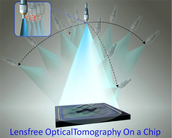
Schematic diagram of the lens-free tomography setup showing the angles of rotation for the light source to illuminate a sample
Ozcan, a researcher at the California NanoSystems Institute at UCLA, makes the analogy that a traditional optical microscope is like a huge set of pipes delivering content, in the form of images, to the user. “Lens-free imaging removes the pipes altogether by utilizing an entirely new design,” he said.
The system takes advantage of the fact that organic structures, such as cells, are partially transparent. So by shining a light on a sample of cells, the shadows created reveal not only the cells’ outlines but details about their sub-cellular structures as well.
“These details can be captured and analyzed if the shadow is directed onto a digital sensor array,” Isikman said. “The end result of this process is an image taken without using a lens.”
Ozcan envisions this lens-free imaging system as one component in a lab-on-a-chip platform. It could potentially fit beneath a microfluidic chip, a tool for the precise control and manipulation of sub-millimeter biological samples and fluids, and the two tools would operate in tandem, with the microfluidic chip depositing and subsequently removing a sample from the lens-free imager in an automated, or high-throughput, process.
We present a lens-free optical tomographic microscope, which enables imaging a large volume of approximately 15 mm3 on a chip, with a spatial resolution of less than 1 micron by 1 micron by 3 microns in x, y and z dimensions, respectively. In this lens-free tomography modality, the sample is placed directly on a digital sensor array with, e.g., less than 4 mm distance to its active area. A partially coherent light source placed approximately 70 mm away from the sensor is employed to record lens-free in-line holograms of the sample from different viewing angles. At each illumination angle, multiple subpixel shifted holograms are also recorded, which are digitally processed using a pixel superresolution technique to create a single high-resolution hologram of each angular projection of the object. These superresolved holograms are digitally reconstructed for an angular range of ± 50°, which are then back-projected to compute tomograms of the sample. In order to minimize the artifacts due to limited angular range of tilted illumination, a dual-axis tomography scheme is adopted, where the light source is rotated along two orthogonal axes. Tomographic imaging performance is quantified using microbeads of different dimensions, as well as by imaging wild-type Caenorhabditis elegans. Probing a large volume with a decent 3D spatial resolution, this lens-free optical tomography platform on a chip could provide a powerful tool for high-throughput imaging applications in, e.g., cell and developmental biology.
11 pages of supplemental material
If you liked this article, please give it a quick review on ycombinator or StumbleUpon. Thanks

Brian Wang is a Futurist Thought Leader and a popular Science blogger with 1 million readers per month. His blog Nextbigfuture.com is ranked #1 Science News Blog. It covers many disruptive technology and trends including Space, Robotics, Artificial Intelligence, Medicine, Anti-aging Biotechnology, and Nanotechnology.
Known for identifying cutting edge technologies, he is currently a Co-Founder of a startup and fundraiser for high potential early-stage companies. He is the Head of Research for Allocations for deep technology investments and an Angel Investor at Space Angels.
A frequent speaker at corporations, he has been a TEDx speaker, a Singularity University speaker and guest at numerous interviews for radio and podcasts. He is open to public speaking and advising engagements.