

University of Pennsylvania has a micron precision cell manipulation system using micromagnets
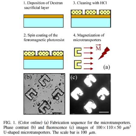
For biomedical applications, such as single cell manipulation, it is important to fabricate microstructures that can be powered and controlled wirelessly in fluidic environments. In this letter, we describe the construction and operation of truly micron-sized, biocompatible ferromagnetic microtransporters driven by external magnetic fields. Microtransporters were fabricated using a simple, single step fabrication method and can be produced in large numbers. We demonstrate that they can be navigated to manipulate single cells with micron-size precision without disturbing the local environment.
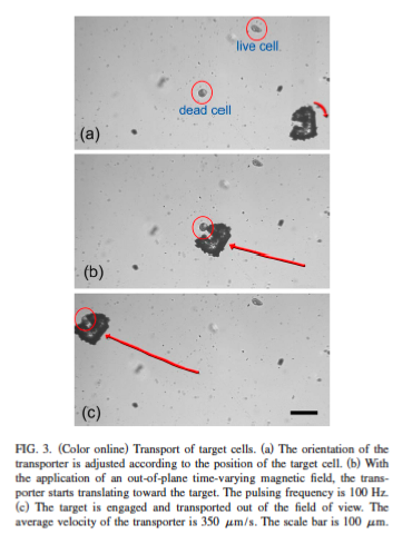
Cells and microtransporters were released into the same open channel. First, a microtransporter/target pair was selected and the center of mass of the target dead cell was calculated using our tracking algorithm. These cells can easily be detected due to their distinct morphological appearance. They have a spherical shape while live cells look like ellipsoids. Next, the orientation of the transporter was adjusted according to the position of the target cell.
In the absence of an out-of-plane magnetic field, the transporter was kept in position while the orientation was changed. By applying a time-varying field with a pulsing frequency of 100 Hz, we were able to induce a smooth motion and translate the transporter toward the target cell and accomplish the engagement. Finally, the cell was cleared out of the field of view. In our experiments, while the manipulation of target cells was performed, the position of other cells kept unchanged unless they were in close proximity (less than 100 microns). This is expected since the flow in the far field falls of inversely with the square of the distance in the low Reynolds regime. We were able to release transported cells and use the same microtransporter multiple times. However, this result cannot be generalized for other cell types.
For applications in which target cells look physically similar to the rest of the cells, fluorescence microscopy can be employed. Target cells can be labeled using fluorescent dyes, fluorescent proteins i.e., GFP or quantum dots. To test the feasibility of our tracking algorithm under fluorescent illumination, we acquired images using several excitation/ emission filter combinations with a 100 W mercury lamp source. The microtransporters were visible with clear contours which make tracking possible.
In order to accomplish the manipulation of cells in a fully automated fashion and estimate the forces applied to the cells by the transporters, we are currently developing an experimentally validated mathematical model describing the dynamics of the system. The behavior of the microtransport-ers depends on their shape, the weight ratio of the suspended magnetite particles, the strength of the applied magnetic fields and the excitation waveform driving the electromagnets.
The effect of each factor needs to be characterized to optimize the overall performance. The microfabrication process can also be improved by measuring the optical properties of the ferromagnetic photoresist and accordingly optimizing
the exposure time.In this letter, a simple method for the fabrication of truly micron-sized, ferromagnetic structures was implemented to fabricate microtransporters. We also demonstrated that single cells can be separated based on their shape without disturbing the local environment. The application area is not limited to cell transport. Positioning cells inside microfluidic channels for single cell analysis is one possibility. We are planning to apply forces locally to specific regions of the cells and examine the cellular reaction to these applied forces.
If you liked this article, please give it a quick review on ycombinator or StumbleUpon. Thanks

Brian Wang is a Futurist Thought Leader and a popular Science blogger with 1 million readers per month. His blog Nextbigfuture.com is ranked #1 Science News Blog. It covers many disruptive technology and trends including Space, Robotics, Artificial Intelligence, Medicine, Anti-aging Biotechnology, and Nanotechnology.
Known for identifying cutting edge technologies, he is currently a Co-Founder of a startup and fundraiser for high potential early-stage companies. He is the Head of Research for Allocations for deep technology investments and an Angel Investor at Space Angels.
A frequent speaker at corporations, he has been a TEDx speaker, a Singularity University speaker and guest at numerous interviews for radio and podcasts. He is open to public speaking and advising engagements.